English



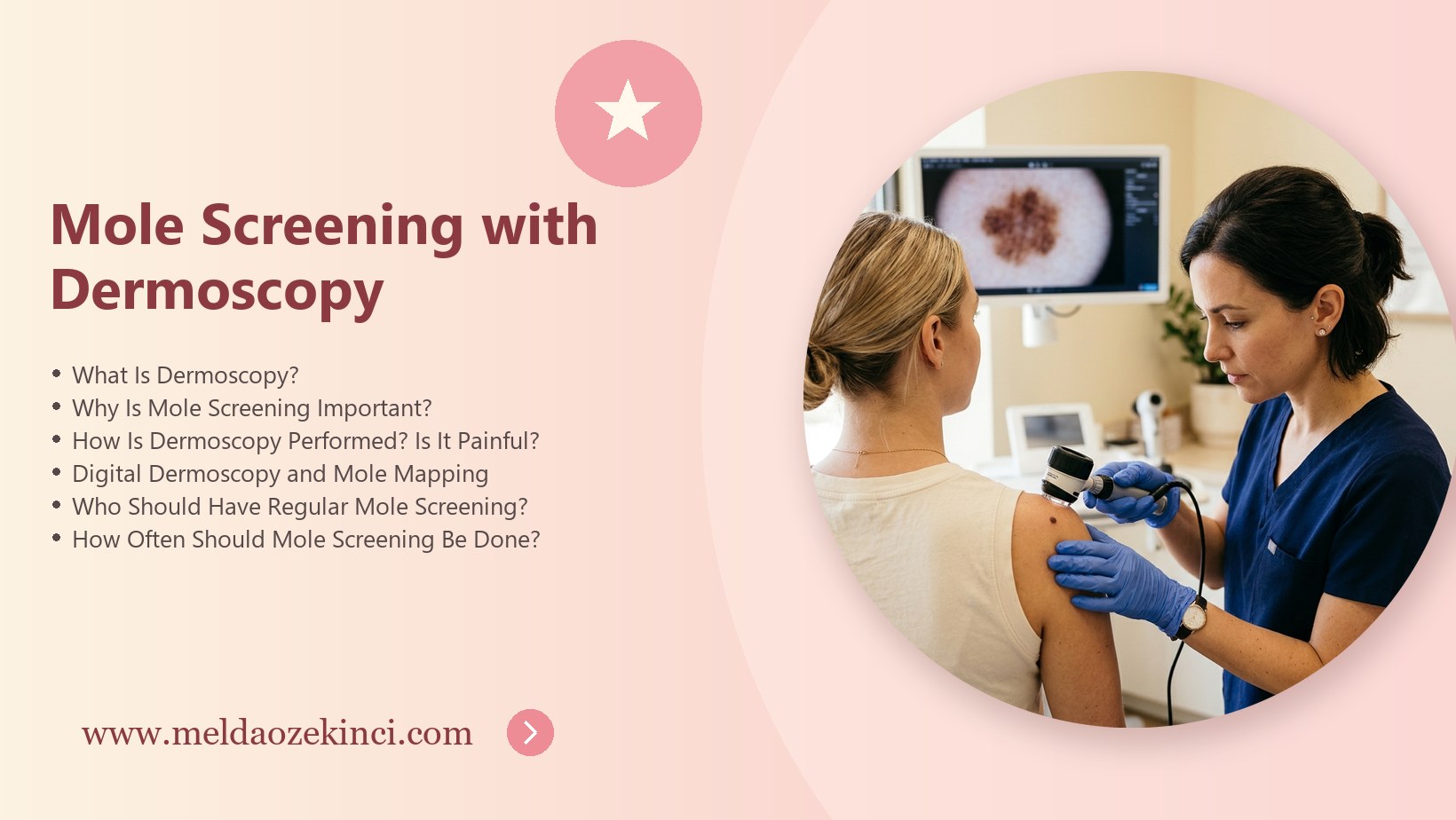

Table of Contents
- What Is Dermoscopy?
- Why Is Mole Screening Important?
- How Is Dermoscopy Performed? Is It Painful?
- Digital Dermoscopy and Mole Mapping
- Who Should Have Regular Mole Screening?
- How Often Should Mole Screening Be Done?
- Checking Moles at Home: The ABCDE Rule
- What Happens If a Suspicious Mole Is Found?
- Mole Screening with Dermoscopy in Antalya
- Frequently Asked Questions
What Is Dermoscopy?
Dermoscopy is a painless, non-invasive examination that uses special light and magnification to examine the skin surface and pigment structures. It allows structures invisible to the naked eye to be evaluated.
It significantly improves diagnostic accuracy in distinguishing benign moles from suspicious ones and reduces unnecessary mole removals.
Dermoscopy is a non-invasive examination method that allows lesions on the skin surface to be magnified and examined under special light. It makes it possible to evaluate color patterns and structural details that cannot be seen with the naked eye. In addition to moles, dermoscopy is used in the differential diagnosis of many skin lesions.
Dermoscopy is used not only to evaluate moles but also to distinguish other skin cancers such as basal cell carcinoma and squamous cell carcinoma, precancerous lesions caused by sun damage and benign skin growths. This helps avoid unnecessary surgery.
During dermoscopy, the doctor may see that lesions that look similar to the naked eye actually have different structures. In experienced hands, this method significantly increases diagnostic accuracy and reduces both unnecessary worry and unnecessary procedures.
Dermoscopic assessment is directly related to the doctor's experience. To interpret subtle differences in mole structure, it is important that the examination be performed by a dermatologist trained in this field. This ensures that risky lesions are not missed and unnecessary procedures are avoided.
Why Is Mole Screening Important?
Skin cancers, especially melanoma, have very high treatment success when caught early. Regular mole screening helps detect new or changing lesions early.
For people living in regions with long sun exposure such as Antalya, regular checks are even more important because of sun damage and related skin lesions.
The main goal of mole screening is to catch skin cancers, especially melanoma, at the earliest stage. Skin cancers detected early have very high treatment success rates. Regular screening is therefore an important preventive health practice for everyone, especially people in risk groups.
Regular mole screening also helps people get to know their own skin better. During examinations with the doctor, they learn which moles should be monitored and which changes are important. This knowledge makes home checks more informed.
Another important benefit of mole screening is raising awareness about sun protection. During the examination, the doctor offers advice on sun protection habits suited to the person's skin type. These recommendations help reduce new mole formation and sun damage.
Having many moles is not in itself a cause for concern, but these people are advised to have their moles monitored regularly. Since tracking every mole individually can be difficult for people with many moles, digital mapping is a particularly useful method for them.
How Is Dermoscopy Performed? Is It Painful?
During the examination, the dermatoscope is placed on the mole and the magnified image is evaluated by the doctor. The procedure is completely painless and involves no needles or incisions.
A full-body check usually takes 15–30 minutes. Removing nail polish and heavy makeup beforehand allows the nails and scalp to be evaluated as well.
During the examination, the doctor places a handheld device called a dermatoscope on the lesion and evaluates its structures under magnification. The procedure is completely painless and requires no preparation or recovery time. The length of the examination depends on the number of moles.
Before the examination, it is recommended not to apply makeup, self-tanner or heavy creams so that moles can be seen clearly. Not wearing nail polish is helpful so that lesions under the nails can also be assessed. The scalp, soles of the feet and spaces between the toes are also part of the examination.
Because the examination is painless and quick, children can also be evaluated with dermoscopy when necessary. If a child has large congenital moles or rapidly changing lesions, regular follow-up at the intervals recommended by the dermatologist is important.
During check-ups, it is important to tell the doctor about any new moles or changing lesions noticed since the previous examination. The patient's own observations guide the doctor to areas that need attention and make the examination more efficient.
Digital Dermoscopy and Mole Mapping
In digital dermoscopy, magnified images of moles are recorded. At later visits, the same mole can be compared and even the smallest change can be detected.
For people with many moles, total body photography (mole mapping) makes it easy to identify new lesions.
Digital dermoscopy allows high-resolution images of moles to be recorded and compared with these images at later check-ups. Mole mapping involves photographing the entire body surface to determine the location of each mole. These methods make it possible to notice even small changes that appear over time.
In digital follow-up, a change in a mole's size, shape or internal structure over time indicates that it needs closer evaluation. This method also prevents unchanged moles from being removed unnecessarily and avoids unnecessary scars on the skin.
The images obtained during mole mapping create a detailed record of the patient's skin. This record can also be used as a reference for check-ups in a different city or with a different doctor, ensuring continuity of follow-up.
Who Should Have Regular Mole Screening?
People with more than 50 moles, atypical (irregular-looking) moles, fair skin that burns easily, a history of severe sunburns in childhood or a family history of melanoma are at higher risk.
People who have had skin cancer before, tanning bed users and immunosuppressed patients should also be checked regularly.
People with many moles, irregular-looking moles, a family history of melanoma, or fair skin that burns easily should have regular mole screening. Having had severe sunburn in childhood and using medicines that suppress the immune system are also among the factors that increase risk.
People who have previously had skin cancer are at higher risk of developing a new one and need regular follow-up. Having used tanning beds is also a factor that increases skin cancer risk and should be reported to the doctor.
During pregnancy, some moles may darken due to hormonal changes. Most of these changes are harmless, but it is recommended that moles that grow noticeably or change shape be evaluated by a dermatologist during pregnancy as well. Dermoscopy can be used safely in this period too.
How Often Should Mole Screening Be Done?
For adults without risk factors, a yearly check is usually sufficient. For high-risk individuals, the doctor may recommend follow-up every 3–6 months.
If you notice a new mole or a change in an existing one between visits, seek an appointment without waiting.
Screening frequency is determined by the doctor according to the person's level of risk. For people without risk factors, an annual check-up may generally be sufficient, while closer follow-up may be recommended for people at high risk. Shorter follow-up intervals may be planned for moles thought to be changing.
Keeping to the check-up schedule is very important for the success of a screening program. Missing planned visits reduces the effectiveness of comparative assessment. If a new or rapidly changing mole is noticed between check-ups, the doctor should be consulted without waiting for the appointment.
Screening intervals may change over time according to the person's age, number of moles and personal or family history of skin cancer. After each check-up, the doctor tells the patient when the next examination should take place and updates the follow-up plan.
Checking Moles at Home: The ABCDE Rule
Checking your whole body in front of a mirror once a month is recommended. The ABCDE rule: A–Asymmetry, B–Irregular border, C–Color variation, D–Diameter larger than 6 mm, E–Evolution in size, shape or color.
Itching, bleeding, crusting or an “ugly duckling” mole that looks clearly different from the others are also warning signs.
The ABCDE rule summarizes the features to watch for in home mole checks: asymmetry, irregular borders, more than one color, a diameter larger than six millimeters and change over time. Noticing one of these features does not necessarily mean cancer, but it is reason enough to see a dermatologist.
Home mole checks can be done once a month in a well-lit room using a full-length mirror and a hand mirror. For hard-to-see areas such as the back and scalp, it helps to ask someone close for assistance. Photographing moles regularly also makes changes easier to notice.
In addition to the ABCDE rule, a mole that looks clearly different from a person's other moles is also considered a warning sign. Lesions that itch, bleed, crust or appear as sores that do not heal should also be shown to a doctor.
What Happens If a Suspicious Mole Is Found?
Moles with suspicious dermoscopic features are either placed under short-term digital monitoring or surgically removed and sent for pathological examination, according to the doctor’s recommendation.
Further treatment is planned if needed based on the pathology result. Early intervention is the most decisive factor in treatment success.
When a suspicious mole is found, the doctor may recommend short-interval follow-up or surgical removal of the lesion. The removed tissue is examined in a pathology laboratory to establish a definitive diagnosis. This is usually a simple, quick outpatient procedure performed under local anesthesia.
The pathology result of a suspicious lesion usually comes back within a few days to a few weeks. Depending on the result, the doctor and patient decide together whether further treatment is needed. Most removed lesions turn out to be benign, but this assessment is necessary for a definitive diagnosis.
When it is decided to remove a mole, following the doctor's wound care advice after the procedure speeds healing and reduces scarring. Keeping the area clean and dry until the stitches are removed and protecting it from the sun after healing are important.
Mole Screening with Dermoscopy in Antalya
In the consultation with dermatologist Dr. Melda Özekinci, moles are examined in detail with dermoscopy, a personal follow-up plan is created according to your risk, and mole removal is planned when necessary.
Antalya is a region exposed to strong sunlight for much of the year, which is an important factor increasing the risk of skin cancer. For people who work outdoors or frequently spend time at the beach and in the sun, regular dermoscopy check-ups are an important step in protecting skin health.
It is important for reliable results that the clinic chosen for mole screening is experienced in dermoscopy and digital follow-up systems. When the doctor explains the findings clearly and plans the next check-up, regular follow-up becomes easier to maintain.
Living in a sunny region requires making sun protection habits part of daily life. High-SPF sunscreen, a hat, sunglasses and staying in the shade at midday, together with regular mole screening, form the most effective protection against skin cancer.
Frequently Asked Questions
Is dermoscopy painful?
No, it is a completely painless examination.
How long does mole screening take?
A full-body check usually takes 15–30 minutes.
Does picking or scratching a mole cause cancer?
Trauma does not turn a mole into cancer, but it can cause bleeding and infection; changing moles must always be examined.
Is mole screening done in children?
Yes, dermoscopic checks are recommended for children with risk factors.