 English


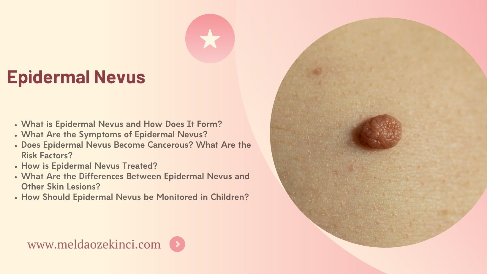

Table of Contents
- What is Epidermal Nevus and How Does It Form?
- What Are the Symptoms of Epidermal Nevus?
- Does Epidermal Nevus Become Cancerous? What Are the Risk Factors?
- How is Epidermal Nevus Treated?
- What Are the Differences Between Epidermal Nevus and Other Skin Lesions?
- How Should Epidermal Nevus be Monitored in Children?
What is Epidermal Nevus and How Does It Form?
Epidermal nevus is a benign skin lesion present from birth that originates from the upper layer of the skin. It usually appears at birth or during childhood. Dermatologist Dr. Melda Özekinci provides important information about the formation process and diagnosis of epidermal nevi.
Epidermal nevi form due to uncontrolled proliferation of skin cells during embryonic development. This results in thickened, raised lesions on the skin, sometimes appearing as lines or patches. The color of the lesions varies between individuals; sometimes skin-colored, sometimes brownish.
According to Dr. Melda Özekinci, these lesions are generally harmless but should be monitored due to aesthetic concerns and rare risk of infection. Treatment options vary depending on the size and extent of the epidermal nevus.
Diagnosis of epidermal nevus is made through dermatological examination. Biopsy may be applied if necessary. Treatment is planned according to the lesion’s size, location, and patient preference. Methods such as laser therapy, surgical excision, or chemical peeling are recommended individually by Dr. Melda Özekinci.
Epidermal nevi are among congenital skin changes and are mostly harmless. However, monitoring and intervention under expert dermatological supervision are important. Dr. Melda Özekinci’s experience makes this process safe and comfortable.
What Are the Symptoms of Epidermal Nevus?
Epidermal nevus are permanent, benign skin lesions visible from birth, prominent on the skin surface. Dermatologist Dr. Melda Özekinci's experience shows that these lesions are usually raised, thick, and brownish or skin-colored. Since symptoms of epidermal nevus can affect quality of life, accurate diagnosis and follow-up are crucial.
These nevi usually appear as linear or patchy patterns on the body and often show unilateral distribution. Lesions may enlarge over time and cause thickening of the skin surface. The most common symptom, thickened lesions, may sometimes cause mild itching or irritation but are mostly painless and cause no significant discomfort.
Dr. Melda Özekinci notes that epidermal nevi may sometimes show crusting, scaling, or color changes. Additionally, increased hair growth or significant thickening of the skin may occur in the lesion area, depending on the type and location of the nevus.
Epidermal nevus is usually noticed in childhood but may sometimes appear in adulthood. Some lesions can become infected or inflamed due to trauma, requiring dermatological intervention and appropriate treatment. Dr. Melda Özekinci emphasizes that early diagnosis and regular monitoring play a critical role in preventing complications.
Also, epidermal nevi should be considered for the rare risk of malignancy. Sudden growth, color change, or bleeding in lesions require expert consultation. Dr. Melda Özekinci stresses that such symptoms must be taken seriously.
Symptoms of epidermal nevus can manifest in various ways, and dermatological evaluation is essential in every case. Regular exams, accurate diagnosis, and appropriate treatment planning using Dr. Melda Özekinci’s expertise help patients achieve the best outcomes.
Does Epidermal Nevus Become Cancerous? What Are the Risk Factors?
Epidermal nevus is a skin condition appearing at birth or in childhood as raised or flat lesions on the skin surface. It is mostly considered harmless but can carry a risk of cancerous transformation in some cases. So, does epidermal nevus become cancerous? The answer depends on the lesion type and monitoring.
Based on the experiences of Dr. Melda Özekinci, epidermal nevi are mostly benign but require regular control and follow-up. Especially rapid growth, color change, or bleeding are risk factors that need expert assessment.
Epidermal nevi usually appear at birth or childhood and often remain unchanged lifelong. However, uncontrolled growth of skin cells may cause a risk of cancer. Though rare, this risk should not be ignored. Long-term irritation, excessive sun exposure, and some genetic factors increase this risk.
Dr. Melda Özekinci recommends regular dermatological follow-up for epidermal nevus. Early detection of lesion changes can prevent cancerous development. Surgical removal may be necessary in some cases.
Most epidermal nevi do not become cancerous, but risk factors should not be overlooked. Professional dermatological control and regular monitoring help prevent complications.
How is Epidermal Nevus Treated?
Epidermal nevus appears as raised, pigmented lesions on the skin from birth or early childhood. They are mostly harmless but may require treatment due to aesthetic concerns or rare complications. Dermatologist Dr. Melda Özekinci is an experienced specialist who customizes the treatment plan based on patient needs.
Several treatment methods can be applied for epidermal nevus. The most common are surgical removal, laser therapy, and cryotherapy. Surgical methods are planned based on lesion size and location and usually provide permanent solutions. However, scar risk is a concern, especially in visible areas like the face.
Laser therapy is a less invasive option for epidermal nevus treatment. Modern laser technologies used at Dr. Melda Özekinci’s clinic effectively reduce lesion color and thickness, speeding healing and minimizing scarring.
Cryotherapy involves freezing and destroying the lesion. This method is preferred for small and superficial epidermal nevi. It is not suitable for every case and requires dermatologist evaluation.
Before starting treatment, Dr. Melda Özekinci conducts a detailed examination and selects the most appropriate method. The treatment plan varies depending on the patient’s age, lesion size, and location. Regular monitoring is critical for treatment success.
What Are the Differences Between Epidermal Nevus and Other Skin Lesions?
Epidermal nevus is a congenital skin problem usually seen as raised, pigmented lesions on the skin surface. So, what are the differences between epidermal nevus and other skin lesions? Dermatologist Dr. Melda Özekinci provides detailed evaluations to offer the most accurate diagnosis and treatment methods to her patients.
Epidermal nevus typically appears from birth and may persist lifelong. Meanwhile, lesions like seborrheic keratosis and lentigo develop mostly with age and have different causes. These differences are important both in appearance and risk factors.
Epidermal nevus is usually irregular and raised, while molluscum contagiosum is smaller, smooth, and a contagious viral infection. Therefore, distinguishing between epidermal nevus and other skin lesions is necessary.
Dr. Melda Özekinci uses advanced techniques like dermoscopy and biopsy when needed for accurate diagnosis. This prevents unnecessary treatments and ensures proper management.
Epidermal nevus has a lower cancer risk compared to other lesions but regular monitoring is important. Knowing differences between skin lesions is critical for early intervention and helps maintain healthy skin.
How Should Epidermal Nevus be Monitored in Children?
Epidermal nevus is common in children, appearing from birth or early childhood. Monitoring these lesions is important to prevent possible complications. Dermatologist Dr. Melda Özekinci emphasizes that the monitoring process in children must be conducted carefully.
In children with epidermal nevus, regular dermatological exams detect early changes in lesion size, shape, and color. Follow-ups at Dr. Melda Özekinci’s clinic are usually every 6 months but may vary based on severity.
Family observation during monitoring is critical. If redness, crusting, bleeding, or rapid growth occurs, immediate dermatological consultation is required because such signs can indicate possible worsening of the epidermal nevus.
Dr. Melda Özekinci notes that advanced imaging methods like dermoscopy are used in epidermal nevus monitoring in children. This allows detailed examination and helps avoid unnecessary interventions.
Although the cancer risk of epidermal nevus is very low, continuous monitoring is essential. Early diagnosis and appropriate intervention protect children’s skin health and improve quality of life.